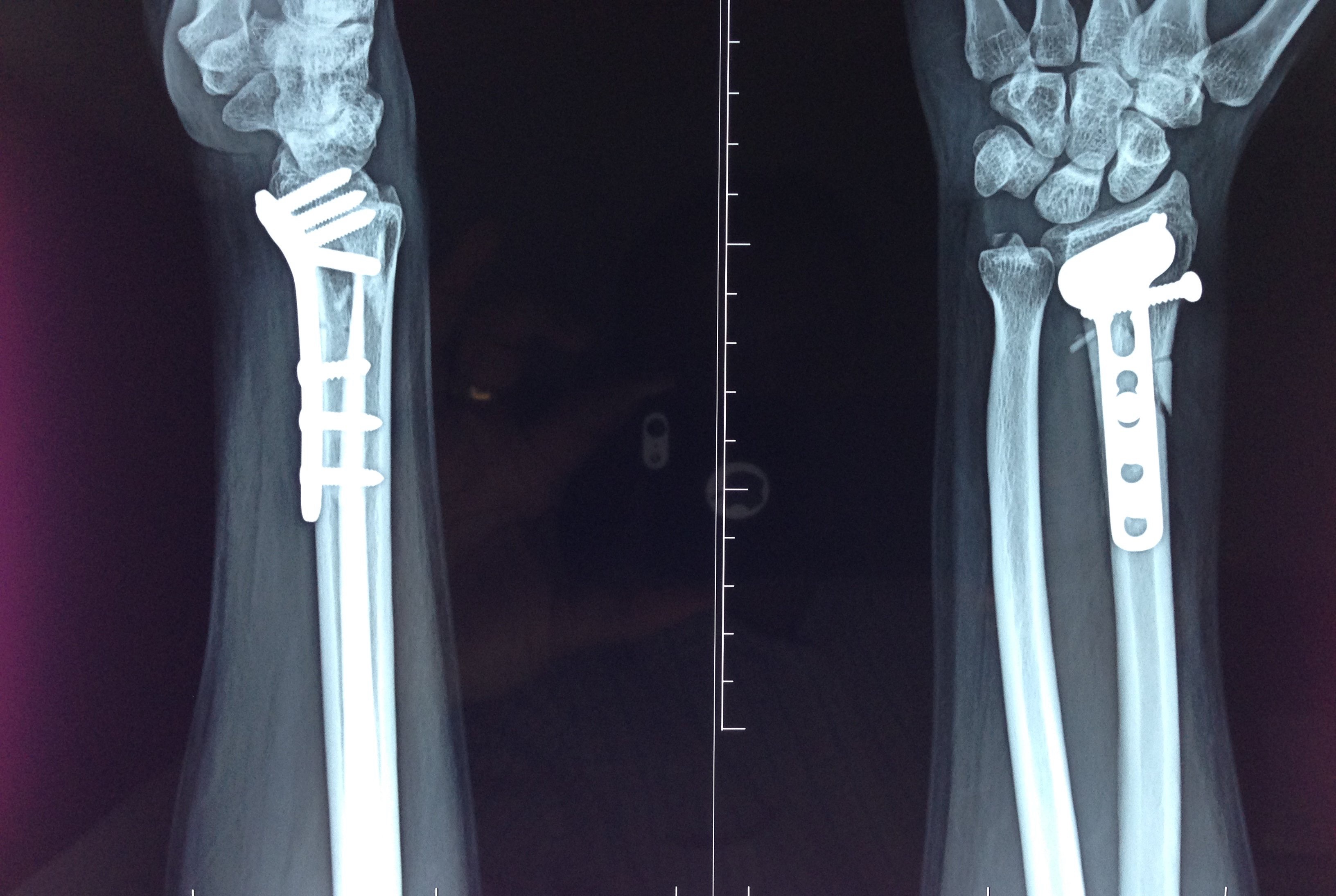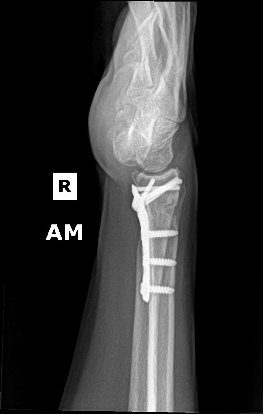

However, other investigators found that displacement of even 1.0 mm resulted in pain and stiffness of wrist. It has been reported that the development of post-traumatic osteoarthrosis in 100% of wrists with articular incongruities of 2.0 mm or more. It has become apparent through the work of several authors that restoration of articular congruency is potentially of greater importance than other criteria.

The wrist and hand are maintained in neutral rotation and held perpendicular to film cassette. For lateral view, the humerus is adducted against the chest wall and elbow is flexed to 90 degrees.

The palm is maintained flat against the cassette. The PA view should be obtained with the humerus abducted 90 degrees from the chest wall, so that the elbow is at the same level as the shoulder and flexed 90 degrees. The routine minimal evaluation for distal radius fractures must include two views-a postero-anterior (PA) view and lateral view. Radiographic imaging is important in diagnosis, classification, treatment and follow-up assessment of these fractures. The degree, direction and extent of the applied load may cause coronal or sagittal splits within the lunate or scaphoid facet. Because of the different areas of bone thickness and density, the fracture patterns tend to propagate between the scaphoid and lunate facets of the distal aspect of the radius. In addition to the extrinsic ligaments of the wrist, the scapholunate interosseous and lunotriquetral interosseous ligaments maintain the scaphoid, lunate and triquetrum in a smooth articular unit that comes into contact with the distal aspect of the radius and the triangular fibrocartilage complex. Only the brachioradialis tendon inserts onto the distal aspect of the radius the other tendons of the wrist pass across the distal aspect of the radius to insert onto the carpal bones or the bases of the metacarpals. The triangular fibrocartilage extends from the rim of the sigmoid notch of the radius to the ulnar styloid process. A central ridge divides the articular surface of the radius into a scaphoid facet and a lunate facet. The dorsal cortical surface of radius thickens to form the Lister tubercle as well as osseous prominences that support the extensors of the wrist in second dorsal compartment. The articular surface of the distal aspect of the radius tilts 21 degrees in the antero-posterior plane and 5 to 11 degrees in the lateral plane. The fracture pattern most commonly observed in geriatric age group is extra-articular while the high-energy intra-articular type is most frequent in young adult patients.Įvery family care physician see this fracture day in and day out and must have a basic knowledge regarding this fracture so as to provide effective treatment when appropriate.Īnatomy of articular interface of distal radius High-energy injuries frequently cause shear and impacted fractures of the articular surface of the distal aspect of the radius with displacement of the fracture fragments. Intra-articular component in distal radius fractures usually signifies high-energy trauma occurring in young adults. It involves an intra-articular fracture of radial styloid of variable size. Chauffer's fracture was described as originally occurring due to backfire of the car starter handles in older models. Reverse Barton's occurs with wrist in palmar-flexion and involves the volar lip. Barton's fracture is the displaced intra-articular coronal plane fracture-subluxation of dorsal lip of the distal radius with displacement of carpus with the fragment. Smith's fractures, also referred to as reverse Colles’ fracture, have palmar tilt of the distal fragment. Colles’ fracture specifically is defined as metaphyseal injury of cortico-cancellous junction (within 2−3 cm of articular surface) of the distal radius with characteristic dorsal tilt, dorsal shift, radial tilt, radial shift, supination and impaction. Abraham Colles is credited with description of the most common fracture pattern affecting distal end radius in 1814, and is classically named after him. They make up 8%−15% of all bony injuries in adults. Distal radius fractures are one of the most common injuries encountered in orthopedic practice.


 0 kommentar(er)
